ملف:HLA-A1.png

من أرابيكا، الموسوعة الحرة
اذهب إلى التنقل اذهب إلى البحث

الملف الأصلي(548 × 609 بكسل حجم الملف: 145 كيلوبايت، نوع MIME: image/png)

ملخص

الوصف
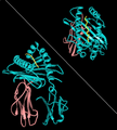
English: Rendering of HLA-A1 with MAGE-1 bound peptide. Two views, from the side showing B2 microglobulin (rose) and HLA-A1 (alpha chain, cyan). Top-right view is looking down through the binding site toward the plasma membrane. The image is derived from PDB:1W72 that was presented in the work:Hulsmeyer et al. (2005) A major histocompatibility complex-peptide-restricted antibody and t cell receptor molecules recognize their target by distinct binding modes: crystal structure of human leukocyte antigen (HLA)-A1-MAGE-A1 in complex with FAB-HYB3. J.Biol.Chem. 280: 2972-2980. Image rendered with PDB ProteinWorkshop 1.50. Several aspects of the image removed for clarity reasons.
المصدر عمل شخصي
المؤلف Pdeitiker

ترخيص

Public domain أنا، مالِك حقوق تأليف ونشر هذا العمل، أجعله في النِّطاق العامِّ، يسري هذا في أرجاء العالم كلِّه.
في بعض البلدان، قد يكون هذا التَّرخيص غيرَ مُمكنٍ قانونيَّاً، في هذه الحالة:
أمنح الجميع حق استخدام هذا العمل لأي غرض دون أي شرط ما لم يفرض القانون شروطًا إضافية.

الشروحات

أضف شرحاً من سطر واحد لما يُمثِّله هذا الملف

العناصر المصورة في هذا الملف

يُصوِّر

تاريخ الملف

اضغط على زمن/تاريخ لرؤية الملف كما بدا في هذا الزمن.

زمن/تاريخصورة مصغرةالأبعادمستخدمتعليق
حالي00:21، 22 أغسطس 2008تصغير للنسخة بتاريخ 00:21، 22 أغسطس 2008548 × 609 (145 كيلوبايت)commonswiki>Pdeitiker{{Information |Description={{en|1=Rendering of HLA-A1 with MAGE-1 bound peptide. Two views, from the side showing B2 microglobulin (rose) and HLA-A1 (alpha chain, cyan). Top-right view is looking down through the binding site toward the plasma membrane. T

ال1 ملف التالي مكررات لهذا الملف (المزيد من التفاصيل):

الصفحة التالية تستخدم هذا الملف: