|
تضامنًا مع حق الشعب الفلسطيني |
ملف:Gray877.png
Gray877.png (378 × 600 بكسل حجم الملف: 36 كيلوبايت، نوع MIME: image/png)
| هذا ملف من ويكيميديا كومنز. معلومات من صفحة وصفه مبينة في الأسفل. كومنز مستودع ملفات ميديا ذو رخصة حرة. |
ملخص
| الوصف |
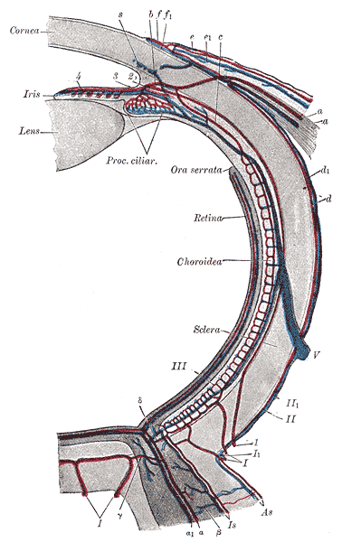
English: Gray's Anatomy plate.
Diagram of the blood vessels of the eye, as seen in a horizontal section. (Leber, after Stöhr.). |
||||||||||||||||||||
| Plate | 877 | ||||||||||||||||||||
| التاريخ | قبل ١٨٥٨ | ||||||||||||||||||||
| المصدر |
|
||||||||||||||||||||
| المؤلف |
|
||||||||||||||||||||
| إصدارات أخرى | File:Gray877 rotated.jpg | ||||||||||||||||||||
.jpg)
كتاب
| هنري غراي: جريز أناتومي
|
|||||||||||||||||||||||
|---|---|---|---|---|---|---|---|---|---|---|---|---|---|---|---|---|---|---|---|---|---|---|---|
| المؤلف |
|
-_Title_page.png) | |||||||||||||||||||||
| المحرر |
Revised by Warren H. Lewis |
||||||||||||||||||||||
| المصور |
|
||||||||||||||||||||||
| العنوان | |||||||||||||||||||||||
| الطبعة |
20 |
||||||||||||||||||||||
| الناشر | |||||||||||||||||||||||
| نوع العمل |
نسخة أو طبعة أو ترجمة |
||||||||||||||||||||||
| نظرة عامة على الصفحة | list of all the plates | ||||||||||||||||||||||
| اللغة |
الإنجليزية |
||||||||||||||||||||||
| تاريخ النشر |
١٩١٨ |
||||||||||||||||||||||
| مكان النّشر |
فيلادلفيا / نيويورك |
||||||||||||||||||||||
| المصدر | Bartleby | ||||||||||||||||||||||

ترخيص
تقع هذه الصُّورة في النِّطاق العامِّ لأَنَّها مَسحٌ مِيكانيكيٌّ أَو ضوئِيٌّ لأَصلٍ يقع في النِّطاق العام أو غير مَحميٍّ بحقوق التَّأليف والنَّشر وليس هُناك أَي دَليلٍ على أَن هُناك حمايةً له ستظهر في وقتٍ لاحِقٍ. إِنَّ الأَصل يقع في النِّطاق العامِّ للسبب التَّالي:
صُمِم هذا الوسم ليُستعمل حيث يُحتمل وجود تحسينات أُضِيفت للأصل، مَثلاً تغيير في السُّطوع أَو التباين أَو مُطابقة الأَلوان، ولكنَّها غير كافية لإِنشاء عملٍ إِبداعيٍّ جديدٍ. يُمكن استخدام هذا الوسم أَيضاً إذا لم يكن هناك مَعلُومات فيما لو أضيفت أي تحسينات أو لا. مِن أَجل المسحات الخام الخالِية مِن أَي تحسينات استعمل وسم {{PD-old}} بدلاً من هذا الوسم، وانظر كومنز:متى يُستعمل وسم النطاق العام الخاص بالصور الممسوحة للمزيد مِن التفاصيل.  | ||||
تاريخ الملف
اضغط على زمن/تاريخ لرؤية الملف كما بدا في هذا الزمن.
| زمن/تاريخ | صورة مصغرة | الأبعاد | مستخدم | تعليق | |
|---|---|---|---|---|---|
| حالي | 00:29، 24 يناير 2007 | | 378 × 600 (36 كيلوبايت) | commonswiki>Pngbot | optimized with optipng |
استخدام الملف
ال1 ملف التالي مكررات لهذا الملف (المزيد من التفاصيل):
- ملف:Gray877.png من ويكيميديا كومنز
ال3 صفحات التالية تستخدم هذا الملف: