ملف:Carboxysome 3 images.png

من أرابيكا، الموسوعة الحرة
اذهب إلى التنقل اذهب إلى البحث

الملف الأصلي(2٬943 × 1٬356 بكسل حجم الملف: 3٫72 ميجابايت، نوع MIME: image/png)

ملخص

الوصف
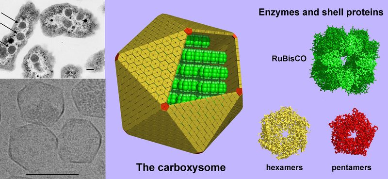
English: (Left, above) A thin-section electron micrograph of H. neapolitanus cells with carboxysomes inside. In one of the cells shown, arrows highlight the visible carboxysomes. (Left, below) Purified carboxysomes (material courtesy of S. Heinhorst and G. Cannon) as visualized by cryo-electron microscopy (courtesy of M. Yeager and K. Dryden). (right) Models for the structure of the carboxysome. Current data suggest that the shell is composed of several hundred hexameric protein building blocks and 12 pentameric building blocks. The three-dimensional atomic structures of the shell proteins have been determined by X-ray crystallography. RuBisCO, the main interior enzyme is shown packed inside in a regular arrangement for simplicity, though the actual organization of the enzymes is not understood yet. The other key enzyme, carbonic anhydrase, which is present in lesser amounts, is not illustrated. Scale bars are 100 nm. (image by T. Yeates).
المصدر Modified version of Image:Carboxysome.png incorporating image taken from Image:Carboxysomes EM.jpg
المؤلف Prof. Todd O. Yeates, UCLA Dept. of Chem. and Biochem.
إصدارات أخرى

[عدل]

SVG:

PNG:


ترخيص

w:ar:مشاع إبداعي
نسب العمل إلى مُؤَلِّفه
يحقُّ لك:
  • مشاركة العمل – نسخ العمل وتوزيعه وبثُّه
  • إعادة إنتاج العمل – تعديل العمل
حسب الشروط التالية:
  • نسب العمل إلى مُؤَلِّفه – يلزم نسب العمل إلى مُؤَلِّفه بشكل مناسب وتوفير رابط للرخصة وتحديد ما إذا أجريت تغييرات. بالإمكان القيام بذلك بأية طريقة معقولة، ولكن ليس بأية طريقة تشير إلى أن المرخِّص يوافقك على الاستعمال.

الشروحات

أضف شرحاً من سطر واحد لما يُمثِّله هذا الملف

العناصر المصورة في هذا الملف

يُصوِّر

ffd0a3efc3aace8e091807d8154f71200484bc5b

٣٬٩٠٥٬٠٠٩ بايت

١٬٣٥٦ بكسل

٢٬٩٤٣ بكسل

تاريخ الملف

اضغط على زمن/تاريخ لرؤية الملف كما بدا في هذا الزمن.

زمن/تاريخصورة مصغرةالأبعادمستخدمتعليق
حالي05:38، 6 أغسطس 2008تصغير للنسخة بتاريخ 05:38، 6 أغسطس 20082٬943 × 1٬356 (3٫72 ميجابايت)commonswiki>TimVickers{{Information |Description={{en|1=(Left, above) A thin-section electron micrograph of H. neapolitanus cells with carboxysomes inside. In one of the cells shown, arrows highlight the visible carboxysomes. (Left, below) Purified carboxysomes (material court

ال1 ملف التالي مكررات لهذا الملف (المزيد من التفاصيل):

الصفحة التالية تستخدم هذا الملف: